

He told them to imagine working in a remote African village at the time of a
disease outbreak, and that all they have at their disposal is a camera cell phone and
an assortment of basic optics lenses and mounts. Would it be possible, Fletcher asked, to convert the camera phone into a sort of mobile microscope that could be used to diagnose the disease?
Using less than US$100 in supplies, they proved that their idea of converting a camera phone into a low-cost, clinical-quality light microscope was feasible.
Now, with funding and software support from Microsoft Research, a small team of graduate, postdoctoral and undergraduate students is working with Fletcher to refine
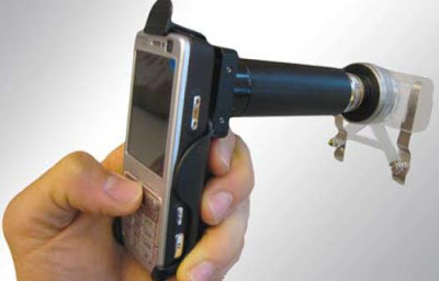
their invention, which they call CellScope.
Cancer patients, who often have to make frequent trips to the hospital for complete blood cell counts, could use a CellScope to do in-home tests and then send the results to their physician. Farmers could use the device to take images of crop blights and then transmit the results to an expert. Fletcher and his team have submitted a patent disclosure on CellScope. But he says the researchers will follow
UC Berkeley’s policy of offering free licenses for technologies used to improve health and welfare in developing countries.
Half the world is middle class and there are over 4 billion cellphones.
Fletcher and his students have developed two lens systems for CellScope so far—a low-magnification scope for viewing conditions such as infections or skin rashes, and a high-magnification scope for analyzing microorganisms in blood, saliva or other samples.
They used a standard clip-on cell-phone holder to make a mounting system for the lenses and for the sample slides. They also have developed an illumination system
using light-emitting diodes (LEDs) powered by the cellphone battery. The team hopes to begin field testing CellScope in early 2009, but first they have to refine the microscope and make it more robust.“Right now, there’s an awful lot of epoxy,” Fletcher says. They are also still working on two software packages: CellScopeCapture, an imaging application that will run on the Microsoft® Windows Mobile® operating system, and CellScopeAnalysis, an image-analyzer.

Brian Wang is a Futurist Thought Leader and a popular Science blogger with 1 million readers per month. His blog Nextbigfuture.com is ranked #1 Science News Blog. It covers many disruptive technology and trends including Space, Robotics, Artificial Intelligence, Medicine, Anti-aging Biotechnology, and Nanotechnology.
Known for identifying cutting edge technologies, he is currently a Co-Founder of a startup and fundraiser for high potential early-stage companies. He is the Head of Research for Allocations for deep technology investments and an Angel Investor at Space Angels.
A frequent speaker at corporations, he has been a TEDx speaker, a Singularity University speaker and guest at numerous interviews for radio and podcasts. He is open to public speaking and advising engagements.