
Ad Support : Nano Technology Netbook Technology News Computer Software

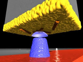
In this diagram, viruses (colored orange) cling to the gold surface (yellow) at the end of a silicon cantilever. A magnetic tip (blue) creates a magnetic field that interacts with the viruses to create an image, using magnetic force resonance microscopy. Image: Martino Poggio, University of Basel
With MRFM, the sample to be examined is attached to the end of a tiny silicon cantilever (about 100 millionths of a meter long and 100 billionths of a meter wide). As a magnetic iron cobalt tip moves close to the sample, the atoms’ nuclear spins become attracted to it and generate a small force on the cantilever. The spins are then repeatedly flipped, causing the cantilever to gently sway back and forth in a synchronous motion. That displacement is measured with a laser beam to create a series of 2-D images of the sample, which are combined to generate a 3-D image.
MRFM resolution is nearly as good (within a factor of 10) of the resolution of electron microscopy, the most sensitive imaging technique that biologists use today. However, unlike electron microscopy, MRFM can image delicate samples like viruses and cells without damaging them
Traditional MRI takes advantage of the very faint magnetic signals emitted by hydrogen nuclei in the sample being imaged. When a powerful magnetic field is applied to the tissue, the nuclei’s magnetic spins align, generating a signal strong enough for an antenna to detect. However, the magnetic spins are so weak that a very large number of atoms (usually more than a trillion) are needed to generate an image, and the best possible resolution is about three millionths of a meter (about half the diameter of a red blood cell). Now scientists are combining the 3-D capability of MRI with the precision of a technique called atomic force microscopy. This combination enables 3-D visualization of tiny specimens such as viruses, cells and potentially structures inside cells — a 100-million-fold improvement over MRI used in hospitals.
Last year, Christian Degen, MIT assistant professor of chemistry, and colleagues at the IBM Almaden Research Center, where Degen worked as a postdoctoral associate before coming to MIT, used that strategy to build the first MRI device that can capture 3-D images of viruses. Last weekend, their paper reporting the ability to take an MRI image of a tobacco mosaic virus was awarded the 2009 Cozzarelli Prize by the National Academy of Sciences, for scientific excellence and originality in the engineering and applied sciences category.
If you liked this article, please give it a quick review on Reddit, or StumbleUpon. Thanks
Supporting Advertising
Business Success
How to Make Money
Executive Jobs
Paid Surveys
Thank You

Brian Wang is a Futurist Thought Leader and a popular Science blogger with 1 million readers per month. His blog Nextbigfuture.com is ranked #1 Science News Blog. It covers many disruptive technology and trends including Space, Robotics, Artificial Intelligence, Medicine, Anti-aging Biotechnology, and Nanotechnology.
Known for identifying cutting edge technologies, he is currently a Co-Founder of a startup and fundraiser for high potential early-stage companies. He is the Head of Research for Allocations for deep technology investments and an Angel Investor at Space Angels.
A frequent speaker at corporations, he has been a TEDx speaker, a Singularity University speaker and guest at numerous interviews for radio and podcasts. He is open to public speaking and advising engagements.