
1. Kurzwelai reports that a European consortium has developed the Megaframe Imager, an ultrafast camera capable of recording images at one million frames per second. It allows for cellular and sub-cellular imaging, neural imaging, biosensing, DNA and protein microarray scanning, automotive collision studies, and high-sensitivity astronomical observations.
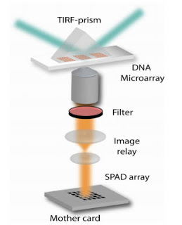
Fluorescence lifetime biosensing with DNA microarrays and a CMOS-SPAD imager

Fluorescence lifetime of dye molecules is a sensitive reporter on local microenvironment which is generally independent of fluorophores concentration and can be used as a means of discrimination between molecules with spectrally overlapping emission. It is therefore a potentially powerful multiplexed detection modality in biosensing but requires extremely low light level operation typical of biological analyte concentrations, long data acquisition periods and on-chip processing capability to realize these advantages. We report here fluorescence lifetime data obtained using a CMOS-SPAD imager in conjunction with DNA microarrays and TIRF excitation geometry. This enables acquisition of single photon arrival time histograms for a 320 pixel FLIM map within less than 26 seconds exposure time. From this, we resolve distinct lifetime signatures corresponding to dye-labelled HCV and quantum-dot-labelled HCMV nucleic acid targets at concentrations as low as 10 nM.
2. Molecular photoacoustic imaging of angiogenesis with integrin-targeted gold nanobeacons
New research in the FASEB Journal shows how combining photoacoustic tomography with gold nanobeacons allows researchers to see blood vessel formation in detail without a microscope
See it for yourself: a new breakthrough in imaging technology using a combination of light and sound will allow health care providers to see microscopic details inside the body
Access to this level of detail potentially eliminates the need for some invasive biopsies, but it also has the potential to help health care providers make diagnoses earlier than ever before—even before symptoms arise.

Although it had been thought acquiring direct images of a hydrogen atom, whose diameter is about one-ten-millionth of a millimeter, was impossible, the team managed the feat with a state-of-the-art “scanning transmission electron microscope” while examining vanadium hydride, a hydrogen storage material.
The microscope scanned an electron beam onto a tiny spot, placed at a theoretically calculated location, on the specimen to enable a detector to catch and film the image of the hydrogen atom as well as the vanadium atom.
If you liked this article, please give it a quick review on ycombinator or StumbleUpon. Thanks
Featured articles
Ocean Floor Gold and Copper
Ocean Floor Mining Company

Brian Wang is a Futurist Thought Leader and a popular Science blogger with 1 million readers per month. His blog Nextbigfuture.com is ranked #1 Science News Blog. It covers many disruptive technology and trends including Space, Robotics, Artificial Intelligence, Medicine, Anti-aging Biotechnology, and Nanotechnology.
Known for identifying cutting edge technologies, he is currently a Co-Founder of a startup and fundraiser for high potential early-stage companies. He is the Head of Research for Allocations for deep technology investments and an Angel Investor at Space Angels.
A frequent speaker at corporations, he has been a TEDx speaker, a Singularity University speaker and guest at numerous interviews for radio and podcasts. He is open to public speaking and advising engagements.