
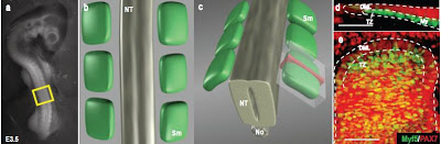

af, Schemes describing how dorsal and transversal views of somites shown throughout the manuscript were acquired at the confocal microscope. ab, general view and scheme of a 3.5 day old chick embryo. c, Confocal images stacks (0.63 μm steps) were acquired through the ectoderm and the entire thickness of somites from immunostained and glycerolclarified embryos (white transparent volume). Dorsal views (e) are projections of stacks of confocal images. Transverse views were made by visualizing the stacks of confocal images at a 90° angle (red volume in c). The thickness of the transverse views was set at 10 μm (d). NT: Neural Tube; Sm: Somite; No: Notochord; DML: Dorso Medial Lip of the dermomyotome; TZ: Transition Zone; My: Myotome.
A team led by developmental biologist Professor Christophe Marcelle has nailed the mechanism that causes stem cells in the embryo to differentiate into specialised cells that form the skeletal muscles of animals’ bodies. The scientists published their results in the British journal Nature
Nature – Neural crest regulates myogenesis through the transient activation of NOTCH

Notch is active during early myogenesis.
How dynamic signalling and extensive tissue rearrangements interact to generate complex patterns and shapes during embryogenesis is poorly understood. Here we characterize the signalling events taking place during early morphogenesis of chick skeletal muscles. We show that muscle progenitors present in somites require the transient activation of NOTCH signalling to undergo terminal differentiation. The NOTCH ligand Delta1 is expressed in a mosaic pattern in neural crest cells that migrate past the somites. Gain and loss of Delta1 function in neural crest modifies NOTCH signalling in somites, which results in delayed or premature myogenesis. Our results indicate that the neural crest regulates early muscle formation by a unique mechanism that relies on the migration of Delta1-expressing neural crest cells to trigger the transient activation of NOTCH signalling in selected muscle progenitors. This dynamic signalling guarantees a balanced and progressive differentiation of the muscle progenitor pool.
Professor Marcelle’s team analysed the differentiation of muscle stem cells in chicken embryos. The mechanisms in birds are identical to those in mammals, so the chick is a good model species for understanding the mechanisms in humans, says team member and the paper’s lead author, Anne Rios.
The scientists investigated the effect of a known signalling pathway called NOTCH on muscle differentiation. They found that differentiation of stem cells to muscle was initiated when NOTCH signalling proteins touched some of the cells. These proteins were carried by passing cells migrating from a different tissue–the neural crest–the progenitor tissue of sensory nerve cells. Muscle formation in the target stem cells occurred only when the NOTCH pathway was triggered briefly by the migrating neural crest cells.
“This kiss-and-run activation of a pathway is a completely novel mechanism of stem cell specification which explains why only some stem cells adopt a muscle cell fate,” Ms Rios said.
Professor Marcelle said that more than 2 per cent of the population was affected by muscle dysfunction. “Muscle frailty in aging and disease imposes a huge economic burden, so it is critical to explore novel avenues of research that could lead to new treatments,” he said.
He said the team would now focus on unraveling the mechanisms of embryonic muscle cell differentiation at the molecular level as a necessary step to regulating regeneration of the muscles in human patients.
13 pages of supplemental information
If you liked this article, please give it a quick review on ycombinator or StumbleUpon. Thanks

Brian Wang is a Futurist Thought Leader and a popular Science blogger with 1 million readers per month. His blog Nextbigfuture.com is ranked #1 Science News Blog. It covers many disruptive technology and trends including Space, Robotics, Artificial Intelligence, Medicine, Anti-aging Biotechnology, and Nanotechnology.
Known for identifying cutting edge technologies, he is currently a Co-Founder of a startup and fundraiser for high potential early-stage companies. He is the Head of Research for Allocations for deep technology investments and an Angel Investor at Space Angels.
A frequent speaker at corporations, he has been a TEDx speaker, a Singularity University speaker and guest at numerous interviews for radio and podcasts. He is open to public speaking and advising engagements.

