
Researchers at Weill Cornell Medical College say they have taken an important step forward in their quest to “turn on” lung regeneration — an advance that could effectively treat millions of people suffering from respiratory disorders.
The research team reports that they have uncovered the biochemical signals in mice that trigger generation of new lung alveoli, the numerous, tiny, grape-like sacs within the lung where oxygen exchange takes place. Specifically, the regenerative signals originate from the specialized endothelial cells that line the interior of blood vessels in the lung.

Highlights
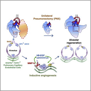
* Pulmonary capillary endothelial cells (PCECs) support alveologenesis
* Autocrine VEGFR2 and FGFR1 activation in PCECs induces MMP14 expression
* MMP14 unmasks EGF receptor ligands, enhancing epithelial cell proliferation
* Injection of activated PCECs or angiocrine factors accelerates lung regeneration
Summary
To identify pathways involved in adult lung regeneration, we employ a unilateral pneumonectomy (PNX) model that promotes regenerative alveolarization in the remaining intact lung. We show that PNX stimulates pulmonary capillary endothelial cells (PCECs) to produce angiocrine growth factors that induce proliferation of epithelial progenitor cells supporting alveologenesis. Endothelial cells trigger expansion of cocultured epithelial cells, forming three-dimensional angiospheres reminiscent of alveolar-capillary sacs. After PNX, endothelial-specific inducible genetic ablation of Vegfr2 and Fgfr1 in mice inhibits production of MMP14, impairing alveolarization. MMP14 promotes expansion of epithelial progenitor cells by unmasking cryptic EGF-like ectodomains that activate the EGF receptor (EGFR). Consistent with this, neutralization of MMP14 impairs EGFR-mediated alveolar regeneration, whereas administration of EGF or intravascular transplantation of MMP14+ PCECs into pneumonectomized Vegfr2/Fgfr1-deficient mice restores alveologenesis and lung inspiratory volume and compliance function. VEGFR2 and FGFR1 activation in PCECs therefore increases MMP14-dependent bioavailability of EGFR ligands to initiate and sustain alveologenesis.
While it has long been known that mice can regenerate and expand the capacity of one lung if the other is missing, this study now identifies molecular triggers behind this process, and the researchers believe these findings are relevant to humans.
“Several adult human organs have the potential upon injury to regenerate to a degree, and while we can readily monitor the pathways involved in the regeneration of liver and bone marrow, it is much more cumbersome to study the regeneration of other adult organs, such as the lung and heart,” says the study’s lead investigator, Dr. Shahin Rafii, who is the Arthur B. Belfer Professor of Genetic Medicine and co-director of the Ansary Stem Cell Institute at Weill Cornell Medical College.
“It is speculated, but not proven, that humans have the potential to regenerate their lung alveoli until they can’t anymore, due to smoking, cancer, or other extensive chronic damage,” says Dr. Rafii, who is also an investigator at the Howard Hughes Medical Institute. “Our hope is to take these findings into the clinic and see if we can induce lung regeneration in patients who need it, such as those with chronic obstructive pulmonary disease (COPD).”
“There is no effective therapy for patients diagnosed with COPD. Based on this study, I envision a day when patients with COPD and other chronic lung diseases may benefit from treatment with factors derived from lung blood vessels that induce lung regeneration,” states Dr. Ronald G. Crystal, who is a co-author of this study and professor of pulmonary and genetic medicine at Weill Cornell.
Dr. Rafii and his researchers had previously uncovered growth factors that control regeneration in the liver and bone marrow, and in both cases, they found that endothelial cells produce the key inductive growth factors, which they defined as “angiocrine factors.” In the current lung study, they discovered the same phenomenon — that blood vessel cells in the lungs jump-start regeneration of alveoli. “Blood vessels are not just the inert plumbing that carries blood. They actively instruct organ regeneration,” says Dr. Rafii. “This is a critical finding. Each organ uses different growth factors within its local vascular system to promote regeneration.”
To conduct this study, Dr. Bi-Sen Ding, a postdoctoral fellow in Dr. Rafii’s lab and the first author of this paper, removed the left lungs of mice and studied the biochemical process of subsequent regeneration of the remaining right lung. Previous pioneering work by Dr. Crystal had shown that when the left lung of mice is removed, the right lung regenerates by 80 percent, effectively replacing most of the lost alveoli. “This regeneration process also restores the physiological respiratory function of the lungs, which is mediated by amplification of various epithelial progenitor cells and regeneration of the alveolar sacs,” says Dr. Ding.
“This regenerative phenomenon, however, only occurs after a trauma that abruptly reduces lung mass. Then the specific subsets of blood vessels in the remaining lung receive a message to start to repopulate alveoli, and our job was to find that signal,” says Dr. Daniel Nolan, a senior scientist in this project who developed methods to characterize the lung blood vessel cells.
The scientists found that removal of the left lung activates receptors on lung endothelial cells that respond to vascular endothelial growth factor (VEGF) and basic fibroblast growth factor (FGF-2). Activation of these receptors promotes the rise of another protein, matrix metalloproteinase-14 (MMP14). The researchers discovered that MMP14, by releasing epidermal growth factors (EGF), initiates the generation of new lung tissue.
When the investigators disabled receptors of VEGF and FGF-2 specifically in the endothelial cells of the mice, the right lung would not regenerate. The defect in the lung regeneration was found to be due to the lack of MMP14 generation from the blood vessels. Remarkably, when these mice received an endothelial cell transplant from a normal mouse, the production of MMP14 was restored, triggering the regeneration of functional alveoli.
“The recovery of lung function and lung mechanics by transplantation of endothelial cells that stimulate MMP14 production may be valuable for designing novel therapies for respiratory disorders,” says Dr. Stefan Worgall, who helped with the functional lung studies in this project. “This study will also help us understand mechanisms for repair in the growing lungs of infants and children,” he adds. Dr. Worgall is associate professor of pediatrics and genetic medicine and distinguished associate professor of pediatric pulmonology.
Given MMP14’s role, Dr. Rafii classifies it as a crucial “angiocrine” signal — a lung endothelial specific growth factor responsible for alveolar regeneration. Dr. Rafii’s team also seeks to reveal the initiation signals resulting in the activation of lung blood vessels. “Changes in local blood flow and biomechanical forces in the remaining lung after removal of the left lung could certainly be one of the initiation cues that induce endothelial activation,” says Dr. Sina Rabbany, who is a co-senior author of this study and a professor of bioengineering at Hofstra University and adjunct associate professor of genetic medicine and bioengineering in medicine at Weill Cornell.
The researchers will next determine if MMP14 and other as-yet unrecognized angiocrine factors are responsible for lung regeneration in humans as well as mice. “We believe the same process goes on in humans, although we have no direct evidence yet,” says Dr. Ding. The study’s authors theorize that patients with COPD (a disorder most often caused by chronic smoking) have so much damage to their lung endothelial cells that they no longer produce the proper inductive signals. “We know smoking damages lungs, but lungs may continue to regenerate alveoli,” says Dr. Koji Shido, a co-author of this study. “But at certain point, significant injury to the endothelial cells could impair their capacity to support lung regeneration.”
“Perhaps replacement of angiocrine factors, or transplantation of normal lung endothelial cells derived from pluripotent stem cells, could restore lung regeneration” speculates Dr. Zev Rosenwaks, who is the director of the Ronald O. Perelman and Claudia Cohen Center for Reproductive Medicine at Weill Cornell, and a co-author of this study. “Currently, we are generating pluripotent stem cells derived from patients with genetic pulmonary disorders to identify potential pathways, which may ultimately enhance our understanding of how lung endothelial cells may improve lung function in these patients.”
If you liked this article, please give it a quick review on ycombinator or StumbleUpon. Thanks

Brian Wang is a Futurist Thought Leader and a popular Science blogger with 1 million readers per month. His blog Nextbigfuture.com is ranked #1 Science News Blog. It covers many disruptive technology and trends including Space, Robotics, Artificial Intelligence, Medicine, Anti-aging Biotechnology, and Nanotechnology.
Known for identifying cutting edge technologies, he is currently a Co-Founder of a startup and fundraiser for high potential early-stage companies. He is the Head of Research for Allocations for deep technology investments and an Angel Investor at Space Angels.
A frequent speaker at corporations, he has been a TEDx speaker, a Singularity University speaker and guest at numerous interviews for radio and podcasts. He is open to public speaking and advising engagements.