
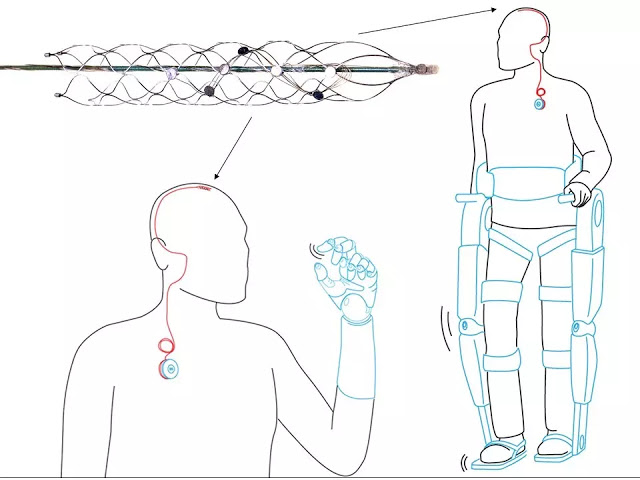
The key to returning mobility is a tiny, matchstick-sized device called a stentrode. It will be implanted into a blood vessel next to the motor cortex, the brain’s control centre – bypassing the need for complex brain surgery.
From there it will pick up brain signals and allow patients to move a robotic exoskeleton attached to their limbs simply by thinking about it.
This notion of wirelessly thought-controlled limbs is within reach, thanks to a collaboration of 39 brilliant minds from 16 departments across the University of Melbourne’s medicine, science, veterinary science and engineering faculties.
The life-changing potential this tiny device offers the ‘Holy Grail’ of bionics.
The stentrode can record brain signals from within a blood vessel. These thoughts are captured, decoded and passed wirelessly through the skin to enable control of an external device, such as a prosthetic limb

Once perfected, the stentrode could also allow U.S. soldiers to move and react faster on the battlefield and should eventually allow them to “talk to” and control futuristic mind-controlled weapons via BMI chips in their brains.

The pre-clinical animal trials of the stentrode, which measures only three millimetres wide, were a success.
The research team demonstrated the stentrode can pick up strong electrical frequencies emitted by the brain that are coded into a computer. The computer then sends a signal to an exoskeleton attached to the arms or legs, enabling movement.

The stentrode device went through hundreds of design changes before researchers were satisfied it met their requirements of being light, flexible, bio-compatible and small enough to be threaded into a one millimetre blood vessel.
It is fitted with tiny recording discs, called electrodes, which sit on the wall of the blood vessel, right next to the brain tissue.
Each disc records electrical activity fired by some 10,000 neurons, which is delivered via delicate wires that run out of the brain, into the neck and emerge into the chest into a wireless transmission system.
The researchers say this transmission can be coded into signals that control an exoskeleton. The first patient will work hard to ‘code’ each of these unique signals to their exoskeleton.
Much like the process of learning to walk or speak again, the process will take many months, until finally, the movement becomes as effortless as driving a car, touch-typing, or writing your name on a form.
“Imagine someone bought a piano for you and you didn’t know how to play,” Dr Oxley explains.
“You know that your hands are physically capable of playing it, but you don’t understand the sequence in which the keys have to be struck. It will take time to use your hands to learn how to play the piano. With our device, you’ve essentially connected an electronic limb to the patient’s brain, but they have to learn how to use it.”
In late 2017, a select group of paralyzed patients from the Royal Melbourne and Austin Hospitals in Australia will be chosen for the trial, where they will be implanted with the stentrode. If the trial succeeds, the technology could become commercially available in as little as six years.
The stentrode could also benefit people with Parkinson’s disease, motor neurone disease, obsessive compulsive disorder and depression and could even predict and manage seizures in epileptic patients.
The stentrode, designed in Melbourne and crafted from a space-age alloy called nitinol, will be inserted into the blood vessel with a catheter fed up through the groin – the same approach that has been used for years for cardiology and removing stroke clots.
When the catheter is inserted into the blood vessel in the brain, it leaves a small cigar-shaped ‘basket’, wired with electrodes, which can record the brainwave activity.
“There is no craniotomy, no risk of infection, it’s all run through the groin and passed inside the body up into the brain,” Professor O’Brien says.
“This has been the Holy Grail for research in bionics – a device that can record brainwave activity over long periods. Inside the blood vessel, it’s protected, it doesn’t damage the brain vessel and can stay there forever.”
Abstract
High-fidelity intracranial electrode arrays for recording and stimulating brain activity have facilitated major advances in the treatment of neurological conditions over the past decade. Traditional arrays require direct implantation into the brain via open craniotomy, which can lead to inflammatory tissue responses, necessitating development of minimally invasive approaches that avoid brain trauma. Here we demonstrate the feasibility of chronically recording brain activity from within a vein using a passive stent-electrode recording array (stentrode). We achieved implantation into a superficial cortical vein overlying the motor cortex via catheter angiography and demonstrate neural recordings in freely moving sheep for up to 190 d. Spectral content and bandwidth of vascular electrocorticography were comparable to those of recordings from epidural surface arrays. Venous internal lumen patency was maintained for the duration of implantation. Stentrodes may have wide ranging applications as a neural interface for treatment of a range of neurological conditions.
SOURCES – University of Melbourne, Youtube, Nature Biotechnology

Brian Wang is a Futurist Thought Leader and a popular Science blogger with 1 million readers per month. His blog Nextbigfuture.com is ranked #1 Science News Blog. It covers many disruptive technology and trends including Space, Robotics, Artificial Intelligence, Medicine, Anti-aging Biotechnology, and Nanotechnology.
Known for identifying cutting edge technologies, he is currently a Co-Founder of a startup and fundraiser for high potential early-stage companies. He is the Head of Research for Allocations for deep technology investments and an Angel Investor at Space Angels.
A frequent speaker at corporations, he has been a TEDx speaker, a Singularity University speaker and guest at numerous interviews for radio and podcasts. He is open to public speaking and advising engagements.