
A new NMR microscope gives researchers an improved instrument to study fundamental physical processes. It also offers new possibilities for medical science, for example to better study proteins in Alzheimer patients’ brains.
This is a major step forward towards the ultimate goal of a device that can measure variations at the nanoscale to study the properties of the inhomogeneous electron systems which are at the forefront of modern condensed-matter physics, such as the surface states of topological insulators, oxide interfaces and other strongly correlated electron systems including the high-Tc superconductors
If you get a knee injury, physicians use an MRI machine to look right through the skin and see what exactly is the problem. For this trick, doctors make use of the fact that our body’s atomic nuclei are electrically charged and spin around their axis. Just like small electromagnets they induce their own magnetic field. By placing the knee in a uniform magnetic field, the nuclei line up with their axis pointing in the same direction. The MRI machine then sends a specific type of radio waves through the knee, causing some axes to flip. After turning off this signal, those nuclei flip back after some time, under excitation of a small radio wave. Those waves give away the atoms’ location, and provide physicians with an accurate image of the knee.
NMR
MRI is the medical application of Nuclear Magnetic Resonance (NMR), which is based on the same principle and was invented by physicists to conduct fundamental research on materials. One of the things they study with NMR is the so-called relaxation time. This is the time scale at which the nuclei flip back and it gives a lot of information about a material’s properties.

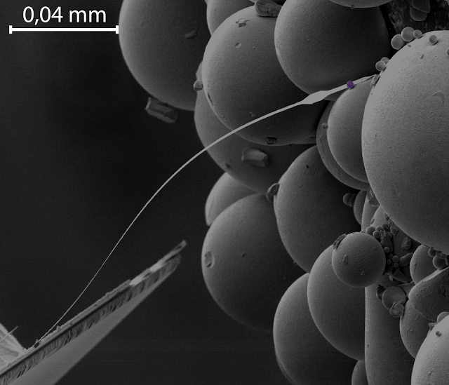
NMR microscope, consisting of a thin wire and a small magnetic ball (fake color purple). The purple ball induces a uniform magnetic field, so that the surrounding atomic nuclei all line up with their axis pointing in the same direction. The researchers send radio waves through their sample, causing some nuclei to flip the other way, and measure how long it takes before they flip back again.
Microscope
To study materials on the smallest of scales as well, physicists go one step further and develop NMR microscopes, with which they study the mechanics behind physical processes at the level of a group of atoms. Now Leiden PhD students Jelmer Wagenaar and Arthur de Haan have built an NMR microscope, together with principal investigator Tjerk Oosterkamp, that operates at a record temperature of 42 milliKelvin—close to absolute zero. In their article in Physical Review Applied they prove it works by measuring the relaxation time of copper. They achieved a thousand times higher sensitivity than existing NMR microscopes—also a world record.
Alzheimer
With their microscope, they give physicists an instrument to conduct fundamental research on many physical phenomena, like systems displaying strange behavior in extreme cold. And like NMR eventually led to MRI machines in hospitals, NMR microscopes have great potential too. Wagenaar: ‘One example is that you might be able to use our technique to study Alzheimer patients’ brains at the molecular level, in order to find out how iron is locked up in proteins.’
Abstract
Nuclear spin-lattice relaxation times are measured on copper using magnetic-resonance force microscopy performed at temperatures down to 42 mK. The low temperature is verified by comparison with the Korringa relation. Measuring spin-lattice relaxation times locally at very low temperatures opens up the possibility to measure the magnetic properties of inhomogeneous electron systems realized in oxide interfaces, topological insulators and other strongly correlated electron systems such as high-Tc superconductors.
SOURCES- Arxiv, Leiden Institute

Brian Wang is a Futurist Thought Leader and a popular Science blogger with 1 million readers per month. His blog Nextbigfuture.com is ranked #1 Science News Blog. It covers many disruptive technology and trends including Space, Robotics, Artificial Intelligence, Medicine, Anti-aging Biotechnology, and Nanotechnology.
Known for identifying cutting edge technologies, he is currently a Co-Founder of a startup and fundraiser for high potential early-stage companies. He is the Head of Research for Allocations for deep technology investments and an Angel Investor at Space Angels.
A frequent speaker at corporations, he has been a TEDx speaker, a Singularity University speaker and guest at numerous interviews for radio and podcasts. He is open to public speaking and advising engagements.