

Nature Materials – To repair complexly shaped tissue defects, an injectable cell carrier is desirable to achieve an accurate fit and to minimize surgical intervention. However, the injectable carriers available at present have limitations, and are not used clinically for cartilage regeneration. Here, we report nanofibrous hollow microspheres self-assembled from star-shaped biodegradable polymers as an injectable cell carrier. The nanofibrous hollow microspheres, integrating the extracellular-matrix-mimicking architecture with a highly porous injectable form, were shown to efficiently accommodate cells and enhance cartilage regeneration, compared with control microspheres. The nanofibrous hollow microspheres also supported a significantly larger amount of, and higher-quality, cartilage regeneration than the chondrocytes-alone group in an ectopic implantation model. In a critical-size rabbit osteochondral defect-repair model, the nanofibrous hollow microspheres/chondrocytes group achieved substantially better cartilage repair than the chondrocytes-alone group that simulates the clinically available autologous chondrocyte implantation procedure. These results indicate that the nanofibrous hollow microspheres are an excellent injectable cell carrier for cartilage regeneration.
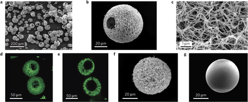
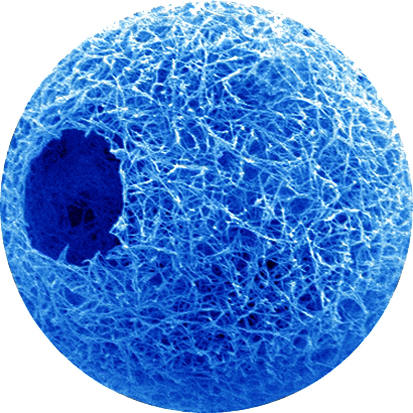
Characterization of nanofibrous hollow microspheres, nanofibrous microspheres and solid-interior microspheres.

1: A schematic of SS-PLLA synthesis and nanofibrous hollow microsphere fabrication. a, PAMAM (G2) as an initiator for the synthesis of SS-PLLA. The colours show the successive PAMAM generations. b, The SS-PLLA synthesized. Red coils represent the PLLA chains. Note that some hydroxyl groups on the PAMAM surface did not react

Schematic illustration of the hollow core formation. In the first step (A), glycerol (purple) was gradually mixed into SS-PLLA solution (aqua), and was a dispersed phase in the emulsification system. As more glycerol was added, there was a phase inversion process, in which SS-PLLA solution became liquid micro-beads surrounded by glycerol (B). Due to its high viscosity, the initially formed glycerol drops were encapsulated in the SS-PLLA microspheres, which were demonstrated in Figure S9. The un-reacted hydroxyl groups (red dots) of the dendrimer cores on the SS-PLLA molecules may serve as a “glycerol-philic” moiety to surround and stabilize the glycerol cores, while the PLLA branches (green) may serve as a “glycerol-phobic” moiety. Similarly, the un-reacted hydroxyl groups (red dots) on the dendrimer cores (blue) of the SS-PLLA molecules may concentrate more around glycerol domains that bridge the outer continuous glycerol phase and the inner glycerol core of the microspheres (C). As the TIPS process proceeded, the PLLA branches further aggregated to form the nanofibers and the glycerol domains remained. After the microspheres were solidified and the encapsulated glycerol in the microspheres and on the shells was extracted, the hollowcores and the open holes on the shells of hollow microsphere formed (D).
Repairing tissue is very difficult and success is extremely limited by a shortage of donor tissue, says Ma, who also has an appointment at the U-M College of Engineering. The procedure gives hope to people with certain types of cartilage injuries for which there aren’t good treatments now. It also provides a better alternative to ACI, which is a clinical method of treating cartilage injuries where the patient’s own cells are directly injected into the patient’s body. The quality of the tissue repair by the ACI technique isn’t good because the cells are injected loosely and are not supported by a carrier that simulates the natural environment for the cells, Ma says.
To repair complex or oddly shaped tissue defects, an injectable cell carrier is desirable to achieve accurate fit and to minimize surgery, he says. Ma’s lab has been working on a biomimetic strategy to design a cell matrix—a system that copies biology and supports the cells as they grow and form tissue—using biodegradable nanofibers.
Ma says the nanofibrous hollow microspheres are highly porous, which allows nutrients to enter easily, and they mimic the functions of cellular matrix in the body. Additionally, the nanofibers in these hollow microspheres do not generate much degradation byproducts that could hurt the cells, he says.
The nanofibrous hollow spheres are combined with cells and then injected into the wound. When the nanofiber spheres, which are slightly bigger than the cells they carry, degrade at the wound site, the cells they are carrying have already gotten a good start growing because the nanofiber spheres provide an environment in which the cells naturally thrive.
This approach has been more successful than the traditional cell matrix currently used in tissue growth, he says. Until now, there has been no way to make such a matrix injectable so it’s not been used to deliver cells to complex-shaped wounds.
During testing, the nanofiber repair group grew as much as three to four times more tissue than the control group, Ma says. The next step is to see how the new cell carrier works in larger animals and eventually in people to repair cartilage and other tissue types.

Hole size of the nanofibrous hollow microspheres is affected by the
polymer concentration. SEM images of nanofibrous hollow microspheres prepared from
SS-PLLA with varying concentrations: (A) 1.0% (wt/v); (B) 6.0% (wt/v).
32 pages of supplemental material
If you liked this article, please give it a quick review on ycombinator or StumbleUpon. Thanks

Brian Wang is a Futurist Thought Leader and a popular Science blogger with 1 million readers per month. His blog Nextbigfuture.com is ranked #1 Science News Blog. It covers many disruptive technology and trends including Space, Robotics, Artificial Intelligence, Medicine, Anti-aging Biotechnology, and Nanotechnology.
Known for identifying cutting edge technologies, he is currently a Co-Founder of a startup and fundraiser for high potential early-stage companies. He is the Head of Research for Allocations for deep technology investments and an Angel Investor at Space Angels.
A frequent speaker at corporations, he has been a TEDx speaker, a Singularity University speaker and guest at numerous interviews for radio and podcasts. He is open to public speaking and advising engagements.