
Medical researchers might realize a range of breakthroughs if they could look deep inside living biological cells, but existing methods for imaging either lack the desired sensitivity and resolution or require conditions that lead to cell death, such as cryogenic temperatures. Recently, however, a team of Harvard University-led researchers working on DARPA’s Quantum-Assisted Sensing and Readout (QuASAR) program demonstrated imaging of magnetic structures inside of living cells. Using equipment operated at room temperature and pressure, the team was able to display detail down to 400 nanometers, which is roughly the size of two measles viruses.

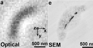
Bright-field image of a magnetotactic bacterium (left) and scanning electron microscope image of the same bacterium (right).
Nature – Optical magnetic imaging of living cells
The researchers used imperfections in diamond known as nitrogen-vacancy (NV) color centers to function as high-precision probes of the magnetic fields produced by living magnetotactic bacteria—organisms that contain magnetic nanoparticles. Using an array of these NV color centers engineered at specific points and density within a diamond chip, the researchers were able to locate the magnetic structures in each bacterium and construct images of the magnetic fields they produced.
The team’s findings have several potential applications and could lead to additional areas of study:
* In principle, this technique would enable detailed, real-time observation of internal cellular processes, such as cell death, evolution and division, and how cells are affected by disease.
* The researchers’ measurements are directly applicable to studying formation of magnetic nanoparticles in other organisms, which is of interest for contrast enhancement in magnetic resonance imaging (MRI), and has been linked to neurodegenerative disorders.
* Formation of magnetic nanoparticles has been proposed as a mechanism for magnetic navigation in higher organisms.
Magnetic imaging is a powerful tool for probing biological and physical systems. However, existing techniques either have poor spatial resolution compared to optical microscopy and are hence not generally applicable to imaging of sub-cellular structure (for example, magnetic resonance imaging), or entail operating conditions that preclude application to living biological samples while providing submicrometre resolution (for example, scanning superconducting quantum interference device microscopy, electron holography and magnetic resonance force microscopy4). Here we demonstrate magnetic imaging of living cells (magnetotactic bacteria) under ambient laboratory conditions and with sub-cellular spatial resolution (400 nanometres), using an optically detected magnetic field imaging array consisting of a nanometre-scale layer of nitrogen–vacancy colour centres implanted at the surface of a diamond chip. With the bacteria placed on the diamond surface, we optically probe the nitrogen–vacancy quantum spin states and rapidly reconstruct images of the vector components of the magnetic field created by chains of magnetic nanoparticles (magnetosomes) produced in the bacteria. We also spatially correlate these magnetic field maps with optical images acquired in the same apparatus. Wide-field microscopy allows parallel optical and magnetic imaging of multiple cells in a population with submicrometre resolution and a field of view in excess of 100 micrometres. Scanning electron microscope images of the bacteria confirm that the correlated optical and magnetic images can be used to locate and characterize the magnetosomes in each bacterium. Our results provide a new capability for imaging bio-magnetic structures in living cells under ambient conditions with high spatial resolution, and will enable the mapping of a wide range of magnetic signals within cells and cellular networks
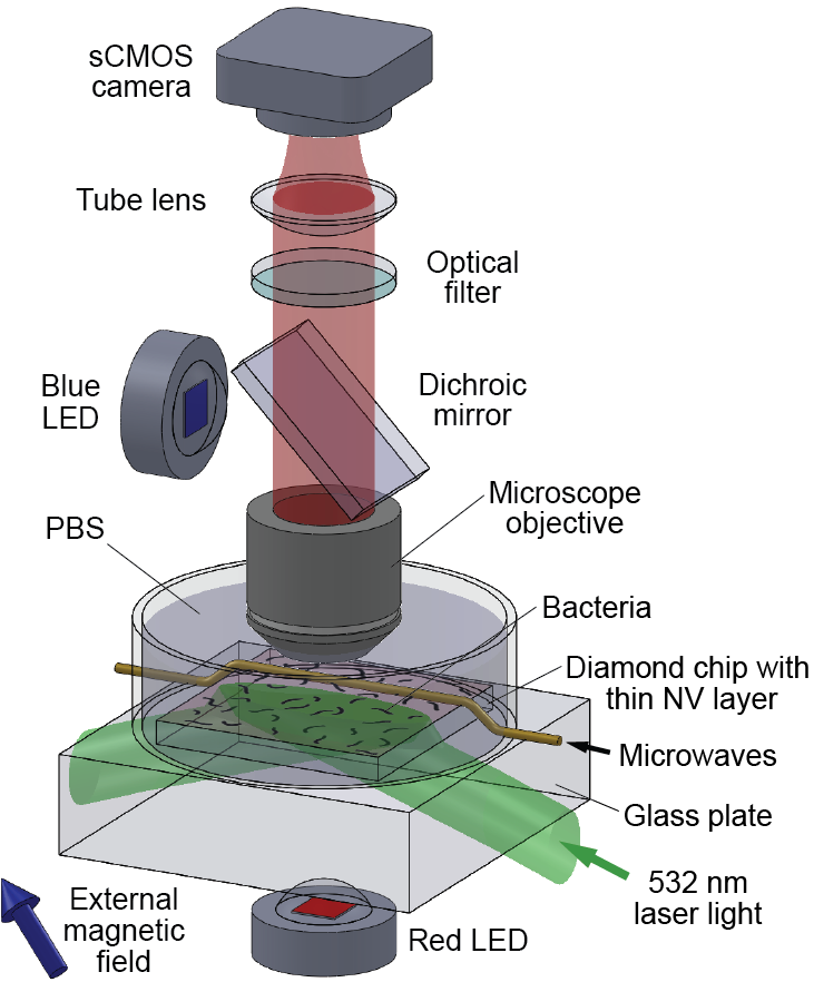
Wide-field fluorescence microscope used for combined optical and magnetic imaging.
If you liked this article, please give it a quick review on ycombinator or StumbleUpon. Thanks

Brian Wang is a Futurist Thought Leader and a popular Science blogger with 1 million readers per month. His blog Nextbigfuture.com is ranked #1 Science News Blog. It covers many disruptive technology and trends including Space, Robotics, Artificial Intelligence, Medicine, Anti-aging Biotechnology, and Nanotechnology.
Known for identifying cutting edge technologies, he is currently a Co-Founder of a startup and fundraiser for high potential early-stage companies. He is the Head of Research for Allocations for deep technology investments and an Angel Investor at Space Angels.
A frequent speaker at corporations, he has been a TEDx speaker, a Singularity University speaker and guest at numerous interviews for radio and podcasts. He is open to public speaking and advising engagements.
