

A DARPA-funded research team has created a novel neural-recording device that can be implanted into the brain through blood vessels, reducing the need for invasive surgery and the risks associated with breaching the blood-brain barrier. The technology was developed under DARPA’s Reliable Neural-Interface Technology (RE-NET) program, and offers new potential for safely expanding the use of brain-machine interfaces (BMIs) to treat physical disabilities and neurological disorders.
In an article published in Nature Biotechnology, researchers in the Vascular Bionics Laboratory at the University of Melbourne led by neurologist Thomas Oxley, M.D., describe proof-of-concept results from a study conducted in sheep that demonstrate high-fidelity measurements taken from the motor cortex—the region of the brain responsible for controlling voluntary movement—using a novel device the size of a small paperclip.
This new device, which Oxley’s team dubbed the “stentrode,” was adapted from off-the-shelf stent technology—a familiar therapeutic tool for clearing and repairing blood vessels—to include an array of electrodes. The researchers also addressed the dual challenge of making the device flexible enough to safely pass through curving blood vessels, yet stiff enough that the array can emerge from the delivery tube at its destination.

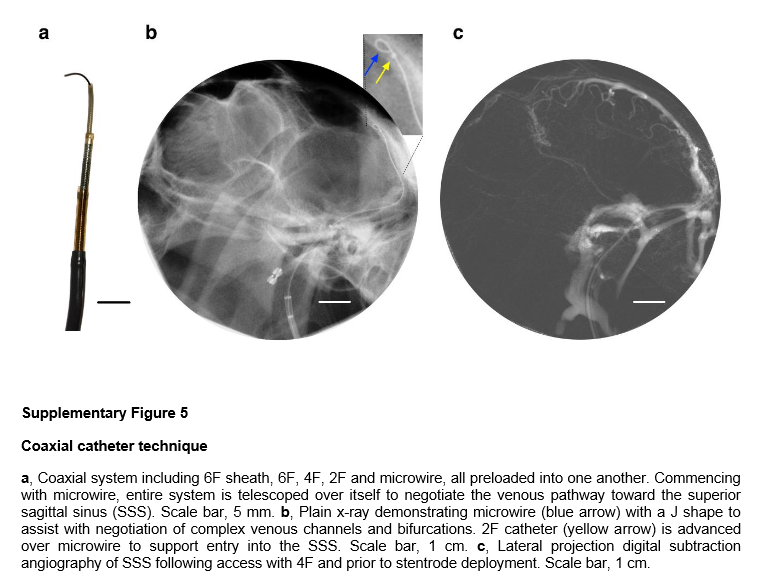
Whereas traditional electrode arrays are implanted into the brain through a surgical procedure that requires opening the skull, the stentrode is delivered via catheter angiography, a much lower-risk procedure. The catheter is inserted into a blood vessel in the neck. Researchers then use real-time imaging to guide the stentrode to a precise location in the brain, where the stentrode then expands and attaches to the walls of the blood vessel to read the activity of nearby neurons. The stentrode technology leverages well-established techniques from the field of endovascular surgery, which uses blood vessels as portals for accessing deep structures while greatly reducing trauma associated with open surgery. Endovascular techniques are routinely used for surgical repair of damaged blood vessels and for installation of devices such as stents and stimulation electrodes for cardiac pacemakers.
For this study, the research team placed the stentrode in a superficial cortical vein overlying the motor cortex, where it could detect electrical signals generated by upper motor neurons in the brain that signal information about movement.
“DARPA has previously demonstrated direct brain control of a prosthetic limb by paralyzed patients fitted with penetrating electrode arrays implanted in the motor cortex during traditional open-brain surgery,” said Doug Weber, the program manager for RE-NET. “By reducing the need for invasive surgery, the stentrode may pave the way for more practical implementations of those kinds of life-changing applications of brain-machine interfaces.”
The published results demonstrate measurement of brain signals with the stentrode that are quantitatively similar to measurements made by commercially available surface electrocorticography arrays implanted during open-brain surgery. Additionally, the study achieved chronic recordings in freely moving sheep for up to 190 days, indicating that implantation of the device could be safe for long-term use.
The research team is planning the first in-human trial of the stentrode in 2017 at the Royal Melbourne Hospital in Melbourne, Australia.
Abstract
High-fidelity intracranial electrode arrays for recording and stimulating brain activity have facilitated major advances in the treatment of neurological conditions over the past decade. Traditional arrays require direct implantation into the brain via open craniotomy, which can lead to inflammatory tissue responses, necessitating development of minimally invasive approaches that avoid brain trauma. Here we demonstrate the feasibility of chronically recording brain activity from within a vein using a passive stent-electrode recording array (stentrode). We achieved implantation into a superficial cortical vein overlying the motor cortex via catheter angiography and demonstrate neural recordings in freely moving sheep for up to 190 d. Spectral content and bandwidth of vascular electrocorticography were comparable to those of recordings from epidural surface arrays. Venous internal lumen patency was maintained for the duration of implantation. Stentrodes may have wide ranging applications as a neural interface for treatment of a range of neurological conditions.
12 pages of supplemental figures
13 pages of supplemental material
SOURCES – DARPA, University of Melbourne, Nature Biotechnology

Brian Wang is a Futurist Thought Leader and a popular Science blogger with 1 million readers per month. His blog Nextbigfuture.com is ranked #1 Science News Blog. It covers many disruptive technology and trends including Space, Robotics, Artificial Intelligence, Medicine, Anti-aging Biotechnology, and Nanotechnology.
Known for identifying cutting edge technologies, he is currently a Co-Founder of a startup and fundraiser for high potential early-stage companies. He is the Head of Research for Allocations for deep technology investments and an Angel Investor at Space Angels.
A frequent speaker at corporations, he has been a TEDx speaker, a Singularity University speaker and guest at numerous interviews for radio and podcasts. He is open to public speaking and advising engagements.