
Researchers have shown the first far-field endoscope based on multimode fiber. Due to its extended depth of field, the holographic endoscope is capable of imaging macroscopic objects in a wide range of distances. They reached the current technological limits, employing the fastest available spatial light modulator and having an unprecedented control over 17 000 fiber modes. This allowed transmitting 0.1 megapixel images, thus reaching the standard definition of modern video endoscopes.
This work should enable semi-rigid minimally invasive multimode fiber probes as a perspective alternative to the rigid endoscopes routinely used in clinical diagnostics and key-hole surgery. Combined with spectroscopic imaging methods the technology has potential for in situ diagnostics at the cellular level.


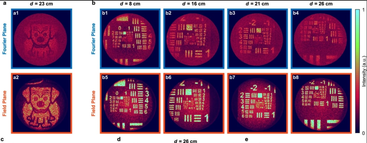
The versatility of the instrument was demonstrated by imaging complex three-dimensional scenes, particularly the interior of a sweet pepper serving as a phantom for biomedically relevant environments, as well as a functioning clockwork mechanism as an example of an object with dynamic complexity. Because of radial k-space conservation of propagating fields in step-index fibers, a larger fraction of the optical power can be directed toward a far-field focus when phase-only modulation is performed in the field plane of the input fiber facet, rather than in its Fourier plane. This allows efficiently manipulating the spatial frequency spectra of the coupled fields and results in far-field imaging with a greatly improved signal-to-noise ratio.
Holographic wavefront manipulation enables converting hair-thin multimode optical fibers into minimally invasive lensless imaging instruments conveying much higher information densities than conventional endoscopes. Their most prominent applications focus on accessing delicate environments, including deep brain compartments, and recording micrometer-scale resolution images of structures in close proximity to the distal end of the instrument. Here, we introduce an alternative “far-field” endoscope capable of imaging macroscopic objects across a large depth of field. The endoscope shaft with dimensions of 0.2 × 0.4 square millimeters consists of two parallel optical fibers: one for illumination and the other for signal collection. The system is optimized for speed, power efficiency, and signal quality, taking into account specific features of light transport through step-index multimode fibers. The characteristics of imaging quality are studied at distances between 20 mm and 400 mm. As a proof-of-concept, we provide imaging inside the cavities of a sweet pepper commonly used as a phantom for biomedically relevant conditions. Furthermore, we test the performance on a functioning mechanical clock, thus verifying its applicability in dynamically changing environments. With the performance reaching the standard definition of video endoscopes, this work paves the way toward the exploitation of minimally invasive holographic micro-endoscopes in clinical and diagnostics applications.
SOURCES – AIP – Observing distant objects with a multimode fiber-based holographic endoscope
Written By Brian Wang, Nextbigfuture.com

Brian Wang is a Futurist Thought Leader and a popular Science blogger with 1 million readers per month. His blog Nextbigfuture.com is ranked #1 Science News Blog. It covers many disruptive technology and trends including Space, Robotics, Artificial Intelligence, Medicine, Anti-aging Biotechnology, and Nanotechnology.
Known for identifying cutting edge technologies, he is currently a Co-Founder of a startup and fundraiser for high potential early-stage companies. He is the Head of Research for Allocations for deep technology investments and an Angel Investor at Space Angels.
A frequent speaker at corporations, he has been a TEDx speaker, a Singularity University speaker and guest at numerous interviews for radio and podcasts. He is open to public speaking and advising engagements.
Test… it appears that comments are working! YAY
Optics look like the laser that powers the interstellar probes, perhaps? Is this related to NASA sending a probe to Uranus to check for gas?
"leave a comment" link should come here. It is currently the same as "Read more" button. Thanx! Also, can there be a button to see only comments, if we have already seen the post? Load as little as possible. Also, the email "View Conversation" button should come here, ideally to this exact spot.
Welcome comments!